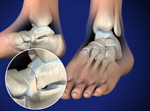

In the present study, we report a surgical treatment of Hawkins type III talar neck fracture through the approach of medial malleolar osteotomy and mini-plate for fixation and discuss the therapeutic effects after long-term follow. This projection is essentially a medial oblique foot with DP tube angulation. Background Fractures of the talar neck are relatively uncommon yet current interventions suffer from a high incidence of complications and poor functional outcomes.
#Talar neck fracture skin#
posterior to the skin margins of the calcaneumĬlear undistorted view of the talus with clear visualization of the talar neck Practical points Final intraoperative fluoroscopy showing a Hawkins 3 talar neck fracture fixed with lateral locking plate, multiple screwings and buried K-wire associated with osteosynthesis of the medial malleolus by double screwing.The knowledge of the blood supply of this bone. The choice of the treatment plan is based on the type of neck fracture and the degree of displacement. Early identification, treatment and rehabilitation are important to avoid long-term complications especially the risk of avascular necrosis. the foot is everted 15° (the same as a medial oblique foot) Talar neck fractures are difficult injuries to treat.the affected leg must be flexed enough that the plantar aspect of the foot is resting on the image receptor.the patient may be supine or upright depending on comfort.The purposes of the present study were to evaluate the rates of early and late complications after operative treatment of talar neck fractures, to ascertain the effect of surgical delay on the development of osteonecrosis, and to determine the functional outcomes after operative treatment. This projection is difficult to perform in the acute setting, however often utilized to assess both the effectiveness of reduction and the monitoring of osteonecrosis 2,3. Background: Talar neck fractures occur infrequently and have been associated with high complication rates. It is particularly useful to assess varus displacement 1,2. This view is specifically indicated when assessing talar neck fracture and/or their follow-up.


 0 kommentar(er)
